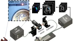
Top:
Schematic of the system.
Botton left:
In vivo imaging of the coronary artery with an implanted NIR fluorescent fibrin-labeled stent using the 4.5F/40MHz hybrid cNIRF-IVUS catheter. Scale bars 1 mm.
Botton right:
In vivo cNIRF-IVUS imaging of inflammation in atherosclerosis. Representative cNIRF-IVUS cross-sectional images are shown in (b) and (c), with lower left magnified insets demonstrating evidence of atherosclerosis in (b). Scale bars 1 mm.
The cNIRF image of inflammatory plaque protease activity (d) correlated with the ex vivo fluorescence reflectance image (FRI) of the resected artery (e).
Figure: CBI
1. Real-time interventional fluorescence molecular imaging
Real-time interventional fluorescence molecular imaging in-particular for surgery and surgical endoscopy in order to improve lesion identification and accurate demarcation of cancer margins. Research is geared toward novel multi-spectral instrumentation and the use of appropriate fluorescent agents with clinical relevance (see Nat. Med. 2011 17:1315-9 and highlights below).
2. Hybrid fluorescence molecular tomography (FMT) and X-ray CT imaging:
The goal of the FMT group is to realize the full potential of the FMT-XCT hybrid imaging system and novel inversion methods using image priors toward accurate quantitative in-vivo molecular imaging in small animals. We further employ FMT-XCT in various preclinical applications to enable iresearch in basic research and drug discovery.
3. Fluorescence and color cryoslicing
Fluorescence and color cryoslicing for accurate measurement of the bio-distribution of fluorescent agents, labeled cells and tissue state. We similarly develop multi-spectral imaging approaches for automatic imaging of tissues during cryoslicing. Current research pursues hardware optimization and the development of imaging methods to correct for photon scattering and deliver images of superior quantification compared to simple photographic fluorescence approaches.
4. Standardization and benchmarking of fluorescence imaging systems.
We investigate the development of algorithms and phantoms for the standardization of fluorescence cameras. This involves validation of performance metrics, illumination correction, and design of benchmarking tests that would eventually allow for high-fidelity fluorescence molecular imaging.
5. Intravascular NIRF imaging
We integrate the development of a cNIRF-IVUS (c=corrected) catheter that enables simultaneous co-registered through-blood imaging of disease related morphological and biological alterations in coronary and peripheral arteries in vivo. We have developed 9F/15MHz peripheral and 4.5F/40MHz coronary hybrid catheters that we tested in swine and rabbit models. A correction algorithm utilizing IVUS information was developed to account for the distance-related fluorescence attenuation due to through-blood imaging.